The Use of 3D Printed Prostate Molds Derived From MRI
By: Dan Sperling, MD
Making three dimensional (3D) objects using a printer has progressed from being an amusing novelty to a readily available and economic research asset. It is especially amazing to think of using a digital file, such as a photograph of a hand or an MRI of an internal organ, to generate a solid replica of the object, layer by layer, on a 3-D printer. A very short video that gives excellent (and fun) examples of 3D printing in action can be found at http://3dprinting.com/what-is-3d-printing/. In it, one person declares, “It turns you from being a passive consumer to an active creator.”
At the National Cancer Institute’s Center for Cancer Research (CCR), 3D printing is used for a multitude of educational and research purposes. “The technology makes the relationship between molecular shape and biological function immediate and tangible, allowing 2D representations of protein, virus or cell structure to ‘jump’ off the page.”[i] While much of the materials and models that result from 3D printing help to illustrate theoretical concepts and processes, CCR’s 3D printers have very practical application for focal therapy of prostate cancer.
Radical (whole-gland) treatments do not require customization; both surgical removal and radiation are simply directed at the entire gland. On the other hand, focal therapy is dependent on accurate identification of tumor location, size and shape in order to tailor the treatment to the patient’s cancer. To determine how well mpMRI can pinpoint the tumor, many studies have been done in which patients who have already chosen prostatectomy undergo mpMRI scans prior to their surgery. For a study of this design, one or more radiologists interpret the images, identifying what they believe to be the tumor(s). After the surgery, their reports are compared with the histopathology (the microscopic examination of individual gland slices) of the surgically removed specimens to determine how well the radiology reports correspond to the histopathology results.
When the correspondence is poor, there are several explanations of why this occurred. One problem concerns the deformable nature of the prostate gland. This means the contours of the gland can be “mushed” because it’s not a hard, rigid object. The gland that was “seen” during the MRI scans may not be exactly the same as the post-prostatectomy specimen. This sets up poor results when the purpose of a clinical trial is to establish a one-to-one anatomical correspondence between imaging and three dimensional specimens. Authors Shah et al. explain:
The correlation between in vivo (in a living organism) MRI images and histopathology is challenging. The prostate is an easily deformable organ, hence, it often deforms during and after prostatectomy. Once the anatomic orientation in the body is lost, it may be difficult to section the prostate in the same plane as the images are obtained. Additionally, in order to improve image quality, prostate MRI is often performed using an endorectal coil, which further deforms the gland.[ii]
Diverse methods have been tried to overcome this problem of matching out-of-anatomic-context 3D specimen slices with two dimensional MRI images. However, thanks to 3D MRI imaging – also called volumetric imaging – the computer can reconstruct a 3D image of a patient’s prostate out of the scan’s successive planes. Converting the virtual 3D image into a solid model provides the key to an elegant matching solution. In August, 2014 the National Cancer Institute reported that staff scientist Marcelino Bernardo was generating such models in order to produce precisely contoured presurgical customized molds that could be used to guide slicing of the same gland after surgical removal, essentially a “forced fit” from the MR image planes to the surgical specimen. According to the NCI Biomedical Informatics Blog
In order to make these customized molds, Bernardo uses open-source software to generate a 3D model of the patient’s prostate. From there, he creates a pre-design shape of a cubic mold and subtracts the prostate model from it in order to generate a file for the mold itself. After that, the mold can be printed on a low-cost 3D printer…[iii]
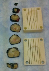
Since the hollow space in the mold is an exact match for the patient’s prostate model, and the imaging planes from the MRI are known, after surgery the prostate specimen can be placed in the mold in the same orientation it occupied in the patient’s body during the imaging, and sliced according to the printed slots that parallel the imaging plane. Below is an image showing slices of a prostate gland next to the two halves of the mold for that prostate, with lines scored for slicing locations:
(NCI image of prostate sections next to 3D-printed mold. Permission applied for.)
For research purposes, the physical specimen slices correspond very accurately with the anatomy revealed by the MRI before surgery. This method overcomes the difficulties of surgery-related deformation and disorientation that may account in some studies for lower correlation results. The 3D-printed prostate mold facilitates validation that mpMRI can accurately identify the location, size and shape of prostate tumors in order to qualify patients for targeted prostate cancer treatment such as the Sperling Prostate Center’s focal laser ablation.
[i] NCI Biomedical Informatics Blog. “Three-Dimensional (3D) Printing: A Gateway to Precision Cancer Medicine.” http://ncip.nci.nih.gov/blog/three-dimensional-3d-printing-gateway-precision-cancer-medicine/
[ii] Shah V, Pohida T, Turkbey B et al. A method for correlating in vivo prostate magnetic resonance imaging and histopathology using indivudalized magnetic resonance-based models. Rev. Sci. Instrum. 80, 104301 (2009); http://dx.doi.org/10.1063/1.3242697
[iii] NCI Biomedical Informatics Blog. “Three-Dimensional (3D) Printing: A Gateway to Precision Cancer Medicine.” http://ncip.nci.nih.gov/blog/three-dimensional-3d-printing-gateway-precision-cancer-medicine/


