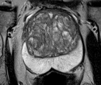
If you look at an MRI image of a prostate gland, what do you see?

You see different shades of gray that form into various useful features that a radiologist reads and interprets. However, a computer is not a human reader. Therefore, in order to be useful for Artificial Intelligence, the measurable physical features must be converted to quantifiable data (quantitative MRI). This data can then be used as descriptors to train Machine Learning software in reading/interpreting the difference between normal prostate tissue and prostate cancer (PCa).
This process is called radiomics, a field that “extracts and mines large number of medical imaging features” that quantify tumor characteristics.[i] Quantifiable MRI parameters include T2 weighted imaging, dynamic contrast enhanced sequences, diffusion weighted imaging, B-values, etc. Their features can then be correlated with actual case records containing clinical information such as patient age, PSA, Gleason grade, tumor stage and genomic reports. Computer developers then train Machine Learning programs to use such clinical factors to predict tumor behavior based on the associations between the quantitative images and real case outcomes. The algorithms in these programs can then calculate probable disease outcomes far more efficiently than humans, and with comparable accuracy. Machine Learning predictions help inform radiologists, quickly pointing the way toward better diagnosis and treatment planning.
A presentation at the 2022 meeting in London of the International Society for Magnetic Resonance in Medicine (ISMRM) described a Machine Learning program that uses a new factor, circulating tumor cells, to predict PCa metastasis (spread). Circulating tumor cells (CTCs) are breakaway cells from the parent tumor that can be detected and counted in a blood draw. They pose a risk of spreading PCa. It’s hard for a lone cancer cell floating around in the blood to overcome the body’s defenses. However, as the parent tumor, or index lesion, continues to grow, it can develop more aggressive cell mutations. If left untreated, there will be more cells released, and they will be more dangerous. Thus, the ever-larger number of circulating tumor cells, and how they have mutated, pose an increasing possibility that some of the cells will colonize other organs or bones, and launch tumors in those locations.
As described in a news story:
The investigators explored any links between radiomic features from multiparametric MRI of the prostate and circulating tumor cell counts in 73 prostate cancer patients. They developed and trained a neural network algorithm to predict circulating tumor cell counts (with a threshold of five or more CTCs indicating risk of metastatic disease). The team conducted 100 training and testing runs of five-fold cross-validation, measuring results using the area under the receiver operating curve (AUC).[ii]
In their population, the investigators found that unfavorable radiomics features correlated with higher CTC levels, predicting a higher probability of PCa spread. The team concluded, “Magnetic resonance imaging radiomics features are promising markers of prostate cancer metastatic risk.”
There is much reason to be optimistic about the branch of Artificial Intelligence (AI) called Machine Learning, and its expanding role in PCa diagnosis and treatment planning. It is very encouraging to know that researchers continue to generate studies confirming that future patients will benefit greatly from AI.
NOTE: This content is solely for purposes of information and does not substitute for diagnostic or medical advice. Talk to your doctor if you are experiencing pelvic pain, or have any other health concerns or questions of a personal medical nature.
References
[i] Parmar, C., Grossmann, P., Bussink, J. et al. Machine Learning methods for Quantitative Radiomic Biomarkers. Sci Rep 5, 13087 (2015).
[ii] Yee, Kate Madden. “MRI radiomics help predict prostate cancer.” Aunt Minnie, May 9, 2022. https://www.auntminnie.com/index.aspx?sec=rca&sub=ismr_2022&pag=dis&ItemID=135768


